
Presentation
Bilateral microphthalmia with decreased visual acuity.
Patient Data
Age: 60 years
Gender: Female
From the case:
Choroidal melanoma
Download
Info
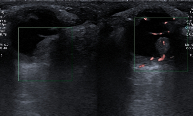
The ultrasound demonstrates:
- small echogenic focus within the surface of the optic nerve head bilaterally with posterior acoustic shadowing in keeping with optic disc drusen
- small echogenic intraocular mass with internal vascularity
From the case:
Choroidal melanoma

Download
Info

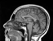
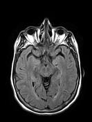
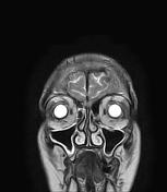
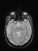
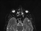
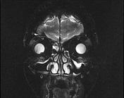
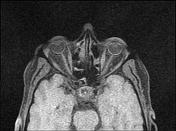
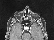
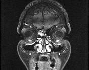
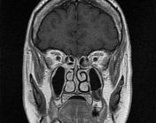
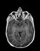
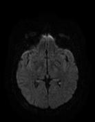
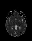
The MRI sequences demonstrate:
- bilateral microphthalmia
- optochiasmatic atrophy
- small right intraocular soft tissue mass at the inferonasal quadrant measuring (7 x 5 x 4 mm) of high signal on T1, low signal on T2 with vivid homogeneous enhancement on postcontrast sequences. No extraocular extension is seen.
- smooth scleral thickening with enhancement choroidal/retinal enhancement
Small vessel chronic ischemic changes are noted.
Case Discussion
MRI features are suggestive of choroidal melanoma in a patient with bilateral microphthalmia with optochiasmatic atrophy. The optic disc drusen was probably an incidental finding.


 Unable to process the form. Check for errors and try again.