
Presentation
Abdominal pain and dyspepsia.
Patient Data


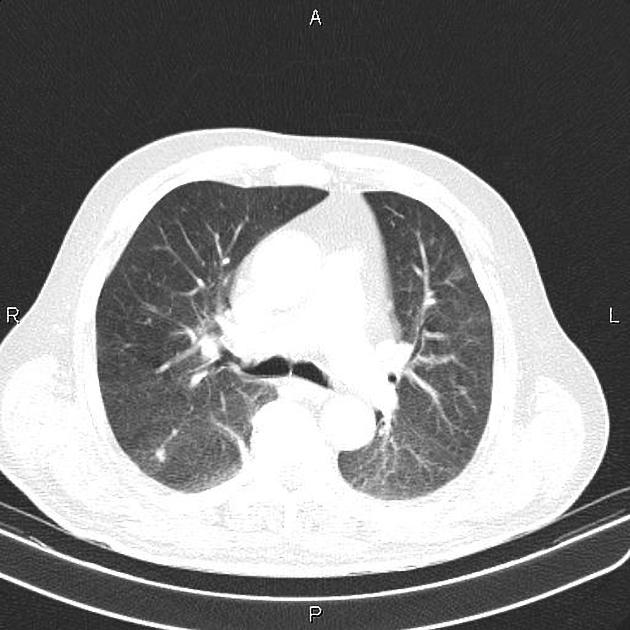
A few nodular opacities are seen at both lungs less than 17 mm. There are also several atelectatic bands scattered bilaterally.
Lymphadenopathy with SAD of 16 mm is noted at subcarina region.
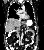
Subtle pleural effusion is noted bilaterally.
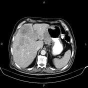
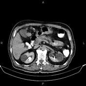
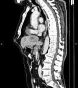
Marked increase wall thickness due to tumoral infiltration is present at gastric lesser curvature accompanied by perigastric fat stranding and several regional lymphadenopathies with maximum SAD of 18 mm. Fat plane between the mass and left liver lobe is obliterated. Additionally, multiple low enhancing masses are observed at liver less than 80 mm.
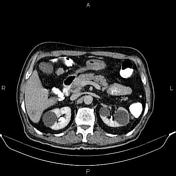
Several non-enhanced simple cortical cysts are seen at both kidneys.
Case Discussion
Pathology proven gastric adenocarcinoma with regional and subcarinal lymphadenopathies; lung and liver metastases.
CT is currently the staging modality of choice because it can help identify the primary tumor, assess for the local spread, and detect nodal involvement and distant metastases


 Unable to process the form. Check for errors and try again.