
Presentation
Febrile convulsions.
Patient Data
Age: 3 months
Gender: Male
From the case:
Normal myelination at 3 months old


Download
Info
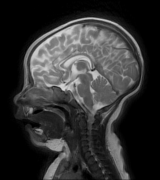
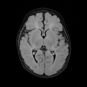
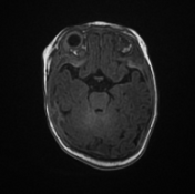
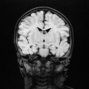
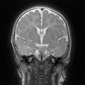
Note the diffuse white matter increased signal on T2 WI, which is normal for the demyelinating white matter in this age group.
Note also the cavum septum pellucidum and prominent extra-axial CSF spaces along the frontal and temporal lobes which are also normal for this age group.
Case Discussion
Normal MRI of the brain at age of 3 months old.


 Unable to process the form. Check for errors and try again.