
Presentation
Right flank pain and hematuria.
Patient Data
Age: 50 years
Gender: Female
From the case:
Renal cell carcinoma


Download
Info

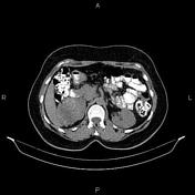
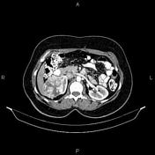
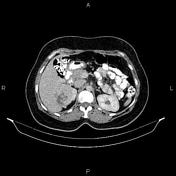
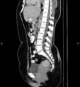
A 75×65 mm partially exophytic heteroenhancing mass is present at upper pole of right kidney. There is no sign of local invasion to adjacent structures and fat plane between the mass and right liver lobe is preserved. No vascular extension and no regional lymphadenopathy are noted.
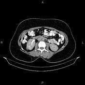
Mild degenerative changes as osteophytosis are seen at the lumbar spine.
Case Discussion
Pathology proven clear cell renal cell carcinoma.
CT is frequently used to both diagnose and stage renal cell carcinomas.


 Unable to process the form. Check for errors and try again.