
Presentation
Left upper and middle abdominal pain for past 3 days, fever. WBC 20K, CRP 200.
Patient Data
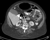


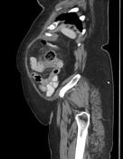

Several diverticula in single loop of jejunum, adjacent to left abdominal wall. One of the diverticula, in the mesenteric wall, measures 3.06 x 3.3 x 3.6 cm and contains fecaloid matter (bezoar?). Marked jejunal wall thickening, hyperemia and prominent mesenteric fat stranding around said diverticulum. No free intra-abdominal gas.
Impression: suspicion of sealed perforation of jejunal diverticulum.
Status post TAVI.
The uterus and adnexa have been removed (TAH-BSO).
Several tiny peripherally located hypodense foci in spleen, too small to characterize.
Few tiny cystic lesions scattered throughout the pancreas, abutting the main pancreatic duct - possibly IPMN.
Several diverticula in sigmoid colon, two of which are calcified.
Anterolisthesis grade I of L3 upon L4. Discogenic changes with vacuum phenomenon in most of intervertebral disks included in the scan; at T12-L1, with Modic type 3 changes.
Summary:
Jejunal diverticulitis, possibly with sealed perforation.

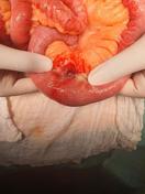
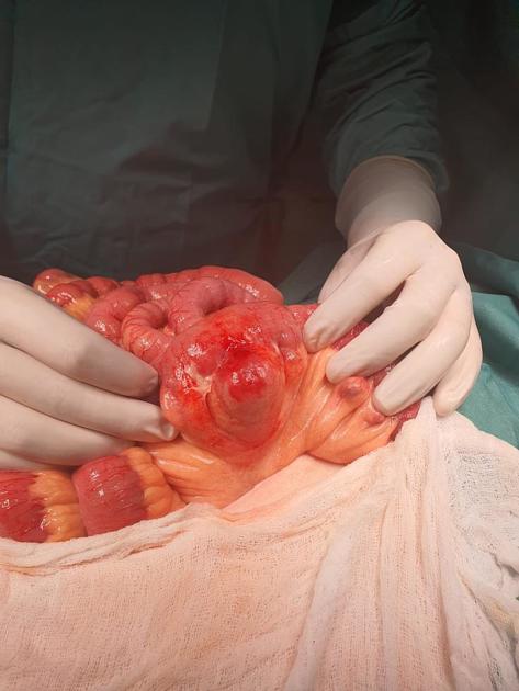
Jejunum showing diverticula and perforation.
Case Discussion
She underwent resection of the diseased part of her jejunum, after which she recovered uneventfully.


 Unable to process the form. Check for errors and try again.