
Recurrent dermatofibrosarcoma protuberans with intracranial extension and chest metastasis
Presentation
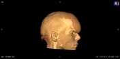
History of surgical excision of forehead mass.
Patient Data


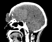
Large forehead soft tissue mass lesion with frontal craniotomy and large intra-cranial extesnion.
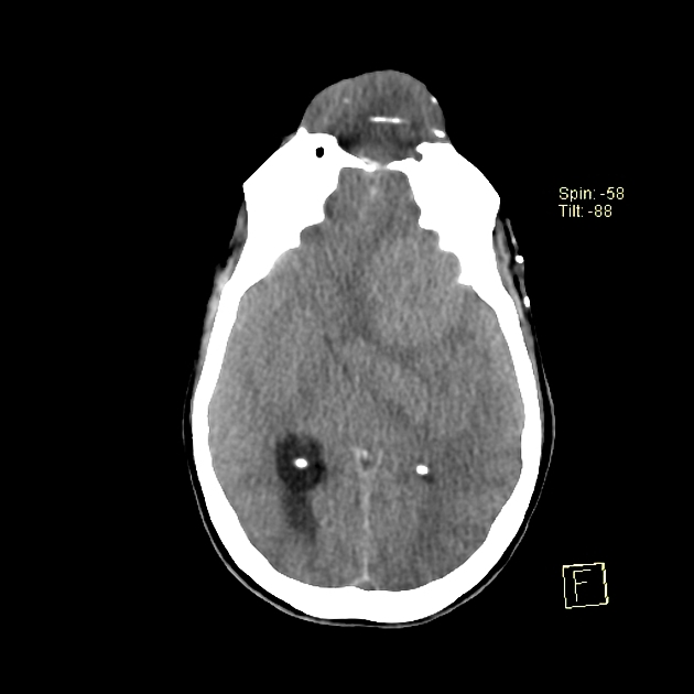
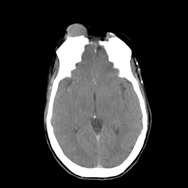
Post-operative
No skin or intracranial masses.
Left frontal craniotomy and adjacent post-surgical change.
Left frontal encephalomalacia.
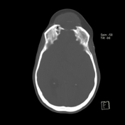
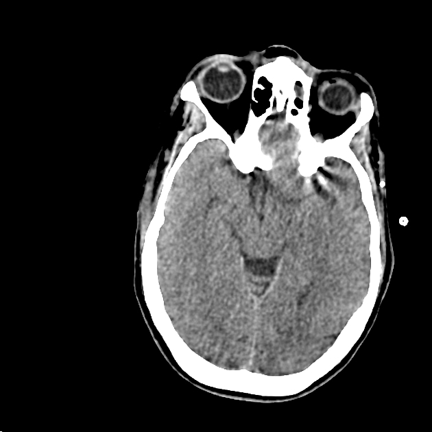
CT 3 months later

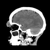
Recurrent forehead and intracranial masses.
CT scan of the chest
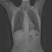
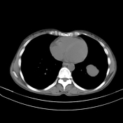
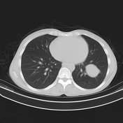
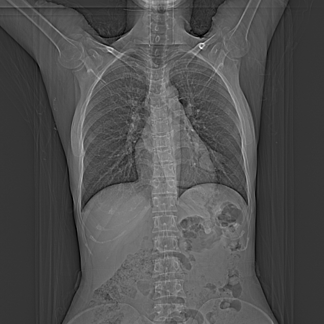
CT scan of the chest showed left lower lobar large lobulated soft tissue mass lesion, likely metastatic.
Case Discussion
This 47-year-old woman had undergone several surgical excisions for forehead dermatofibrosarcoma protuebrans. CT revealed recurrent forehead mass extending via the craniotomy flap into the left frontal extra-axial region. CT scan of the chest revealed left lower lobar pulmonary mass, possibly metastatic.
Dermatofibrosarcoma protuberans (DFSP) is a low-grade malignant tumor arising from dermal and subcutaneous tissues and is the most common cutaneous sarcoma.
Dermatofibrosarcoma protuberans usually have an excellent prognosis after complete surgical resection but have a marked propensity to recur locally if inadequate surgical resection margins are obtained. Metastases are rare, yet if occurred are most commonly to the lungs.
Dermatofibrosarcoma protuebrans are usually fixed to the skin but not involving the underlying structures. However, recurrent or long-standing tumors may invade the underlying fascia, striated muscle, periosteum, and bone 1.
The imaging features of DFSP are non specific. On CT, they appear isoattenuated to hypoattenuated lesions without calcifications and usually uniform if the lesions are small, while heterogeneous in large lesions 2.


 Unable to process the form. Check for errors and try again.