Presentation
Work up for cough and breath shortness.
Patient Data


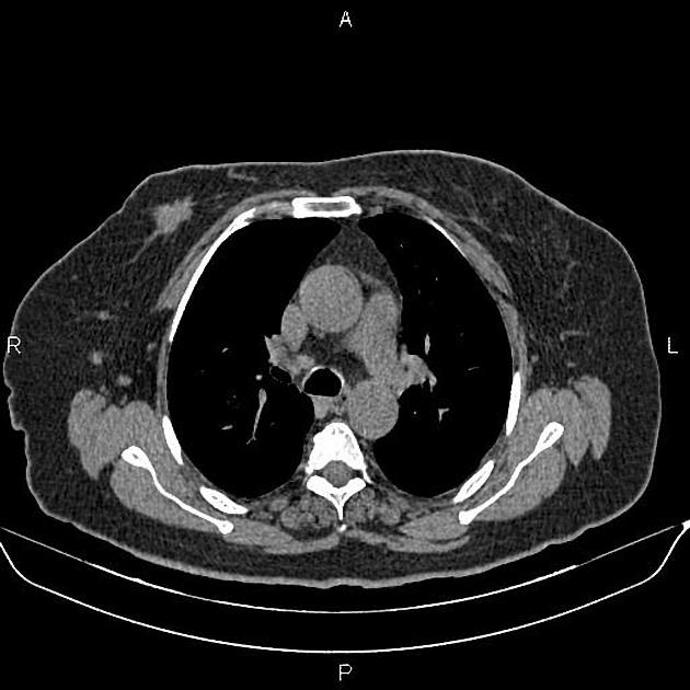
A 27×18 mm mass with irregular margin is present at medial aspect of right breast suggestive for malignant lesion.
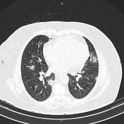
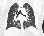
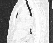
Patchy ill defined, mostly subpleural, ground glass opacities and focal consolidations are seen at both lungs highly suggestive for COVID-19 pneumonia.
In addition, several nodules are seen at both lung fields, less than 10 mm inferring metastases.
Case Discussion
The patient underwent targeted ultrasonography and ultrasound guided core needle biopsy and the histopathology evaluation confirms invasive ductal carcinoma.
This patient had positive RT-PCR testing for COVID-19.


 Unable to process the form. Check for errors and try again.