Q: What is the definition of a volvulus of the colon?
show answer
A: Volvulus is when torsion of the bowel results in obstruction. Volvulus of the colon is when portions of the colon get entangled upon a mesenteric axis, which can cause partial or complete obstruction of the bowel lumen or impair the blood supply. This condition occurs when a redundant and loose mesentery twists around an axis.
Q: Where are the locations of colonic volvulus?
show answer
A: The sigmoid colon is a common location of large bowel torsion, followed by the cecum, transverse colon, and splenic flexure.
Q: What are the general considerations about cecal volvulus?
show answer
A: Cecal volvulus is the torsion of the cecum around its mesentery, which accounts for 10% of intestinal volvuluses. Usually, it occurs in patients 30-60 years old. Congenital anomalies of the right colon fixation to the posterior abdominal usually are present. Pregnancy and recent colonoscopy are factors that result in dilatation of the right colon and may predispose patients to cecal volvulus. Other medical history includes prior abdominal surgery, pelvic mass, adynamic ileus, atonia of the colon, chronic constipation, distant colon obstruction, high fiber intake, extreme exertion, violent coughing, and unpressurized air travel. The size of the cecum at risk for perforation ranges from 9 to 12 cm.
Q: What are the types of cecal?
show answer
A: The types of cecal volvulus are: - type 1, occurs in half of the patients, which forms by a clockwise or counterclockwise axial twisting of the cecum, rotating along with its long axis, located in the right lower quadrant; - type 2, the loop type of cecal volvulus develops from torsion and inversion of a portion of the cecum and of the terminal ileum, and in this type, the cecum gets displaced to an ectopic location, typically occupying the left upper quadrant of the abdomen; - type 3, also known as cecal bascule, which occurs when the cecum folds anteriorly on itself without twisting. It appears as a dilated loop in the mid-abdomen. The cecal bascule is a variant of cecal volvulus. This entity can also lead to bowel ischemia and perforation.
Q: Which are the clinical manifestations of cecal volvulus?
show answer
A: Common clinical symptoms of cecal volvulus include nausea, vomiting, abdominal pain, colicky abdominal pain, acute abdominal pain, obstipation, abdominal distention, and a tympanic abdomen, which may simulate a small-bowel obstruction. Cecal distention will lead to increase wall tension and, without intervention, will progress to ischemia and necrosis.
Q: What are the complications of cecal volvulus?
show answer
A: The complications of cecal volvulus are closed-loop obstruction with vascular ischemia, which can lead to gangrene, perforation, and death.
Q: What are the radiographic features of the cecal volvulus?
show answer
A: Conventional radiography usually reveals dilation of the bowel extending from the right lower quadrant to the left upper quadrant of the abdomen and distal colon decompression. The distended cecum may be displaced anywhere in the abdomen, and it may resemble a coffee bean (the coffee bean sign). The recognition of displacement of the cecum out of the right lower quadrant is the key to diagnosis. It is essential to recognize findings of ischemia in the cecum, which include pneumatosis in the cecal wall, pneumoperitoneum, and portal venous gas. The “bird beak sign” is an essential finding detected during a barium enema.
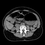
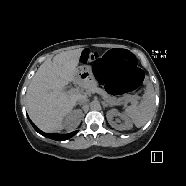
Q: What are the CT findings of the cecal volvulus?
show answer
A: CT findings of cecal volvulus include distension of the cecum, and an abnormal location of the cecal apex and the ileocecal valve, usually in the mid-abdomen or left upper quadrant. Other signs of cecal volvulus include cecal haustral markings, small-bowel distention, distal colon decompression, the bird beak sign, the whirl sign, the ileocecal twist, crossing transition points, which is the X-marks-the-spot, and the split wall sign, that is an invagination of the mesenteric fat that gives the impression of a split in a single twisted loop of bowel. CT findings of ischemia due to cecal volvulus are thickening and hypoenhancement of the wall, pneumatosis intestinalis, portal venous gas, mesenteric stranding, and free air or peritoneal fluid cavity.
Q: How is the treatment of the cecal volvulus?
show answer
A: The options for treatment include endoscopic decompression, cecopexy, cecostomy, manual detorsion, or a right hemicolectomy. Laparoscopic surgery is usually preferred to open surgery. CT findings may change patient treatment by revealing the signs of bowel ischemia, including thickening of the bowel wall, mesenteric hemorrhage, and pneumatosis intestinalis.
Q: Which are the differential diagnosis of cecal volvulus?
show answer
A: The differential diagnosis of cecal volvulus is with bowel obstruction, constipation, pseudo-obstruction, acute mesenteric ischemia, inflammatory bowel disease, irritable bowel syndrome, peptic ulcer disease, appendicitis, sigmoid diverticular disease, and abdominal hernias.


 Unable to process the form. Check for errors and try again.