
Presentation
Abdominal pain and jaundice.
Patient Data


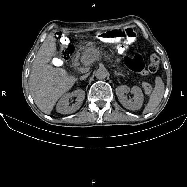
Post-operative changes are seen due to gastrojejunostomy.
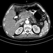
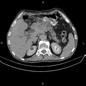
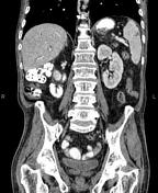
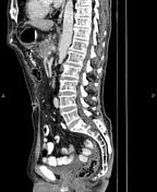
A 48×25 mm cystic solid mass is present in pancreatic neck and proximal of body which encases celiac trunk and proximal portion of portal vein; causing marked distal pancreatic duct dilatation. Additionally, intra and extra hepatic bile ducts are dilated. Peripancreatic fat stranding is evident. The mass is in close contact with gastric antrum and fat plane is infiltrated.
An 8 mm small cyst is present at segment IVa of the liver. Multiple hypervascular masses are also seen at liver less than 38 mm.
A 32 mm non enhanced area is evident at anterior aspect of spleen. Most likely due to post-surgical traumatic lesion.
Several lymphadenopathies are noted at peripancreatic regions.
A little amount free fluid is present at pelvis.
Case Discussion
Pancreatic mass, pathology proven adenocarcinoma, with vascular encasement, local invasion to gastric antrum, regional lymphadenopathies and hepatic metastasis.


 Unable to process the form. Check for errors and try again.