
Presentation
Right flank pain and hematuria.
Patient Data
Age: 60 years
Gender: Female
From the case:
Renal cell carcinoma


Download
Info
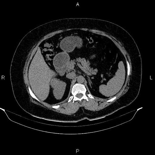
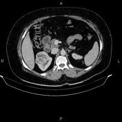
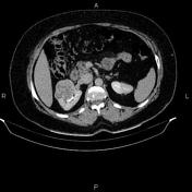
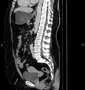
An iso dense mass, 62mm in diameter was seen in upper and mid poles of right kidney. After IV contrast media, it enhances less than normal parenchyma. The mass bulged into renal sinus and compresses pelvicaliceal system.
A 41 mm cyst with a smooth and thin wall, sharp and distinct margination, and a homogenous water density was seen in left kidney. After IV contrast media, it does not enhance and has no discernible wall thickness.
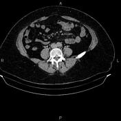
Two small exophytic masses were seen in the uterus inferring fibroids.
Case Discussion
Right renal mass, pathology proven renal cell carcinoma.
No vascular involvement, no regional lymphadenopathy, no metastasis.


 Unable to process the form. Check for errors and try again.