
From the case:
Meckel cave meningioma


Download
Info

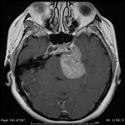
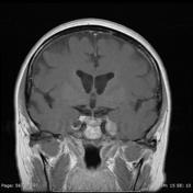
A T1 intermediate and mildly T2 hyperintense mass is centered near the petrous apex of the left temporal bone extending prepontine and exerting mass effect on the left. Post-contrast images show avid enhancement. Features consistent with a left Meckel's cave meningioma.
Case Discussion
See also : tumors of meckel's cave
Histology
Paraffin sections show a moderately hypercellular meningioma with a well developed syncytial architecture. Tumor cells have regular nuclear features. A very occasional mitotic figure is identified. No necrosis is seen and there is no evidence of brain invasion.


 Unable to process the form. Check for errors and try again.