
Presentation
Breath shortness and cough.
Patient Data


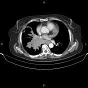
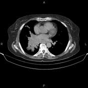
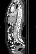

A 65×48 mm mass is present at right lung hilum that encases hilar structures and causing distal parenchymal collapse. Additionally, several nodules are scattered at right lung less than 15 mm.
Multiple mediastinal and right hilar lymphadenopathies are present with SAD less than 28 mm.
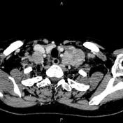
Several lymphadenopathies are present at bilateral lower neck and supraclavicular spaces with maximum SAD of 35 mm.
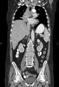
Multiple small hypodense lesions are seen at liver less than 30 mm. Some of them show delayed enhancement.
The gallbladder is not seen at anatomical location due to prior resection.
A 25 mm nodule is present at left adrenal gland.
Case Discussion
Right lung mass; pathology proven small cell carcinoma with mediastinal, hilar, supraclavicular and cervical lymphadenopathies; hepatic and left adrenal metastases.


 Unable to process the form. Check for errors and try again.