
Presentation
Work up for abdominal pain.
Patient Data
Age: 75 years
Gender: Male
From the case:
Hepatic and pelvic hydatid cysts
Download
Info
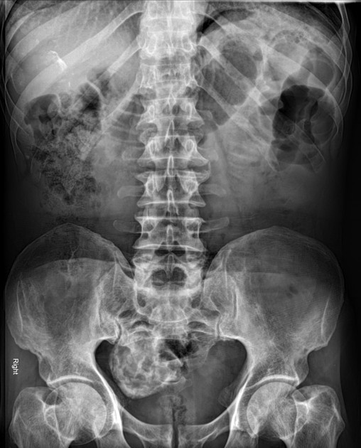
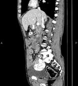
Calcified mass like lesions are seen at right upper quadrant (right liver lobe) as well as right side of the pelvis.
From the case:
Hepatic and pelvic hydatid cysts


Download
Info
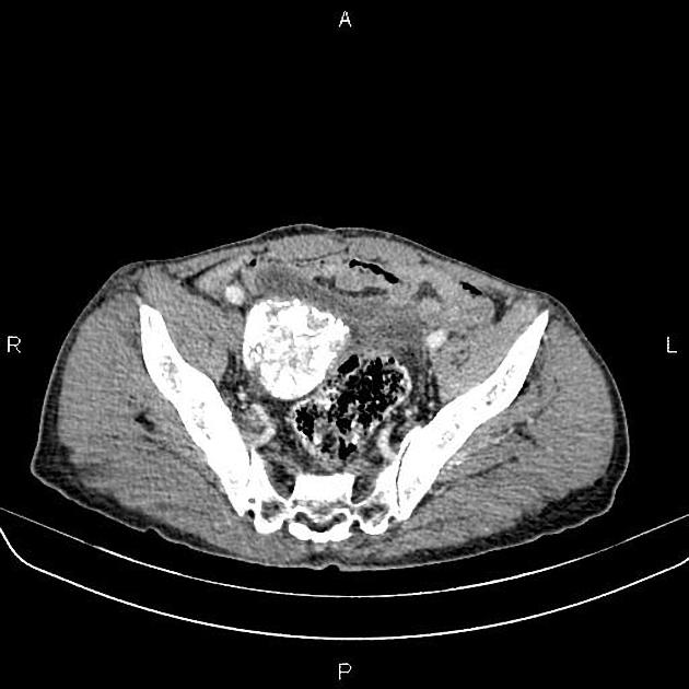
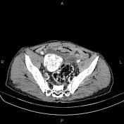
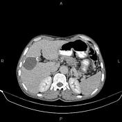
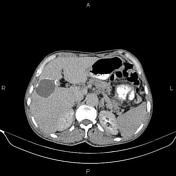
A 52 mm cystic lesion with marginal calcification is present at right liver lobe compatible with hydatid cyst (type IIC). Additionally, a 62 mm calcified hydatid cyst (type III) is noted at right side of the pelvis.
The prostate gland is enlarged.
Case Discussion
Hepatic and pelvic calcified hydatid cysts (type IIC and III).
Based on morphology the cyst can be classified into four different types, see: hydatid cyst classification.


 Unable to process the form. Check for errors and try again.