
Presentation
Abdominal pain.
Patient Data
Age: 80 years
Gender: Female
From the case:
Choledocholithiasis


Download
Info
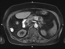
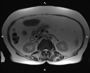
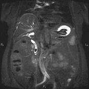
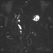
The gallbladder is not seen at anatomical location due to prior resection.
Intra and extra hepatic bile ducts are dilated and CBD measured 17 mm in caliber. An 18 mm low signal filling defect is evident at distal of CBD inferring stone.
Case Discussion
Choledocholithiasis in a 80 years female with abdominal pain.


 Unable to process the form. Check for errors and try again.