
Presentation
Abdominal pain and dyspepsia.
Patient Data
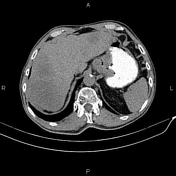

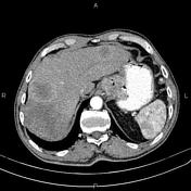
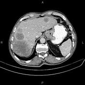
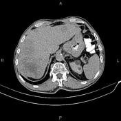
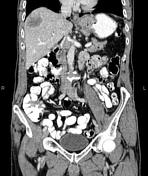

Increased wall thickness because of tumoral infiltration is present at gastric cardia and sub cardia accompanied by a few regional lymphadenopathies with SAD less than 28 mm. Lymphadenopathy with SAD of 26 mm is also noted at upper paracaval regions.
A few small calcified foci are seen at the right liver lobe parenchyma most consistent with healed granuloma. There are also multiple ill defined hetero enhancing masses at liver less than 80 mm.
Several non-enhanced simple cortical cysts are seen in both kidneys.
The prostate gland is enlarged.
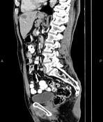
Degenerative changes as osteophytosis are seen at the thoracolumbar spine.
Grade I spondylolisthesis of L5 on S1 is present with bilateral spondylolysis.
Case Discussion
Gastric mass, pathology proven adenocarcinoma, with regional and paracaval lymphadenopathies and hepatic metastases.


 Unable to process the form. Check for errors and try again.