
Presentation
Left pelvic pain.
Patient Data
Age: 25 years
Gender: Male
From the case:
Ewing sarcoma of the pelvis


Download
Info
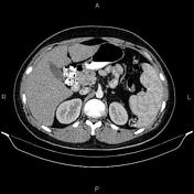
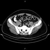
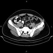
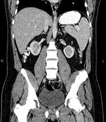
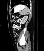
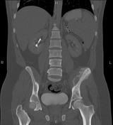
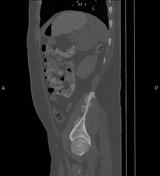
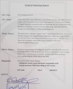
The left iliac bone shows increased density and marked periosteal reaction accompanied by anterior and posterior adjacent soft tissue infiltration. The left sacroiliac joint is intact.
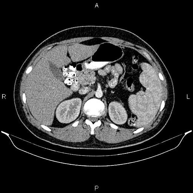
The spleen is enlarged and its cephalocaudal height measured 140 mm.
From the case:
Ewing sarcoma of the pelvis
Download
Info

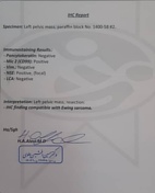
Histopathologic evaluation and IHC confirms malignant small round cell tumor compatible with Ewing sarcoma.
Case Discussion
CT features of an osteosclerotic tumor involves the left iliac wing with large soft tissue component, pathologically proven as an Ewing sarcoma.
Ewing sarcoma typically occurs in children and adolescents between 10 and 20 years of age (95% between 4 and 25 years of age).


 Unable to process the form. Check for errors and try again.