
Presentation
Abdominal pain and dyspepsia.
Patient Data
Age: 65 years
Gender: Male
From the case:
Metastatic gastric cancer


Download
Info
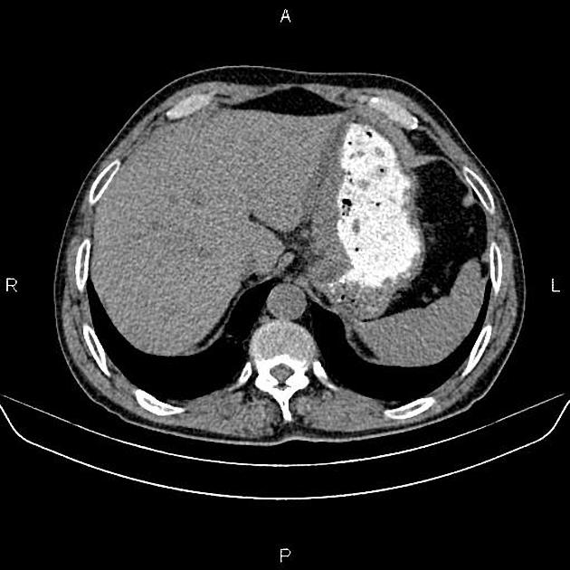
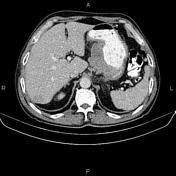
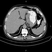
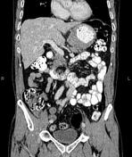
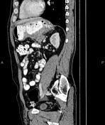
Marked increased wall thickness up to 26 mm due to tumoral infiltration is present at gastric lesser curvature accompanied by perigastric fat stranding and several regional lymphadenopathies with SAD less than 12 mm. There is no sign of local invasion to adjacent structures.
Multiple ill-defined low enhancing masses are seen in the liver less than 20 mm.
The prostate gland is enlarged.
Case Discussion
Gastric mass - pathology proven adenocarcinoma with regional lymphadenopathies and hepatic metastasis.
CT is currently the staging modality of choice because it can help identify the primary tumor, assess for the local spread, and detect nodal involvement and distant metastases.


 Unable to process the form. Check for errors and try again.