
Presentation
Abdominal pain and dyspepsia.
Patient Data


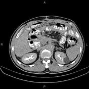
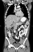
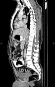
Increased wall thickness due to tumoral infiltration is present at esophagogastric junction, gastric cardia, subcardia and body. Several enlarged perigastric and mesenteric lymphadenopathies are observed.
A little amount ascites and omental thickening are also evident.
Inhomogeneously, the hepatic attenuation value is less than of the spleen, suggesting uneven fatty liver.
A few non-enhanced simple cortical cysts are seen at both kidneys.
The prostate gland is enlarged.
Small fat containing left inguinal hernia is present.
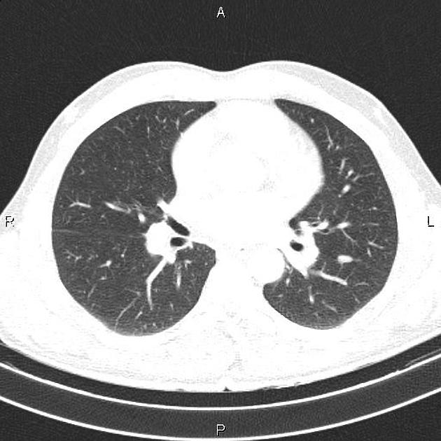
An 11 mm nodule is present on left major fissure most likely fibrotic nodule.
Case Discussion
Esophagogastric mass; pathology proven adenocarcinoma with perigastric and mesenteric lymphadenopathies, ascites and omental thickening.
CT is currently the staging modality of choice because it can help identify the primary tumor, assess for the local spread, and detect nodal involvement and distant metastases.


 Unable to process the form. Check for errors and try again.