
Presentation
Lower abdominal pain, nausea.
Patient Data


Medially coursing appendix of normal width, fills up with contrast material and gas, no periappendiceal fat stranding.
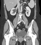
Moderate right hydronephrosis due to small ureteric calculus at level of L3.
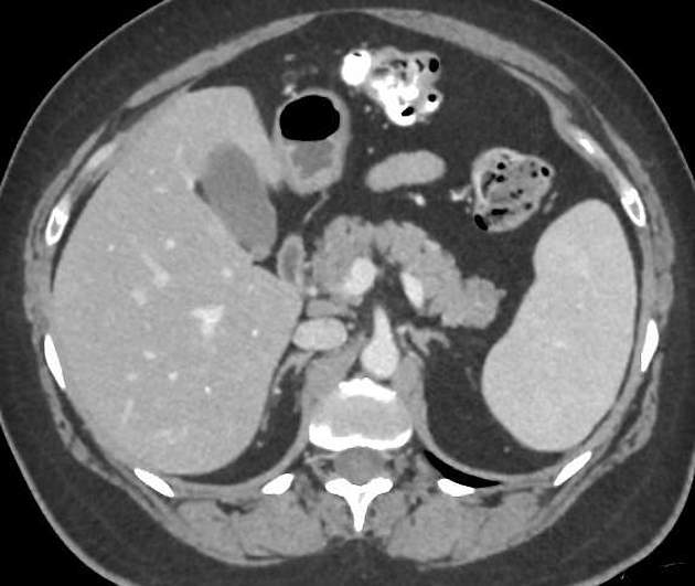
Milk of calcium cyst in lower pole of right kidney (the hyperdense content is dependent, proved by prone CT stone protocol done 2 years earlier). Small parapelvic cyst in upper pole of left kidney.
Incidentally noted retroaortic left renal vein. The left adrenal vein and left ovarian vein drain into a common trunk which then drains into the inferior vena cava (IVC).
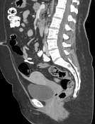
A cord emanates from the uterine fundus in the midline, is isodense to the fundus (i.e. is composed of the fundal external layers), and courses cranially in the direction of the umbilicus - adhesional traction due to post cesarean section?
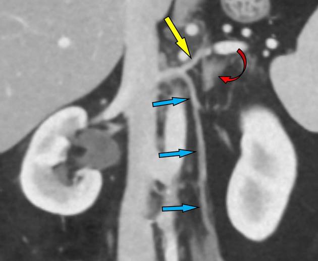
- yellow arrow - left adrenal vein
- red curved arrow - left adrenal
- light blue arrows - left ovarian vein
Case Discussion
Presented with constant lower abdominal pain that started several hours earlier, and nausea. No vomiting, fever, diarrhea or dysuria. History of cesarean section and renal calculi.
Calculus seen in right ureter. She went on to have a cystoscopy with double J insertion.
Incidental rare venous variant where the left adrenal vein and left ovarian vein, both of which usually drain into the left renal vein (retroaortic in the case), drain here into a common trunk (or perhaps the left ovarian vein drains into the left adrenal vein) which drains into the IVC.


 Unable to process the form. Check for errors and try again.