Presentation
Left shoulder pain and asymmetry. ? Sprengle shoulder.
Patient Data
![](https://prod-images-static.radiopaedia.org/images/382556/ba13594ae76909cbeafebc0990b1c1_thumb.jpeg)
![](https://prod-images-static.radiopaedia.org/images/382555/b916a91f38f59492c30df6d7f085f7_thumb.jpeg)
![](https://prod-images-static.radiopaedia.org/images/382557/99e7a3c17b3271c98e9c267ce4f18c_thumb.jpeg)
![](https://prod-images-static.radiopaedia.org/images/382556/ba13594ae76909cbeafebc0990b1c1_big_gallery.jpeg)
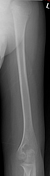
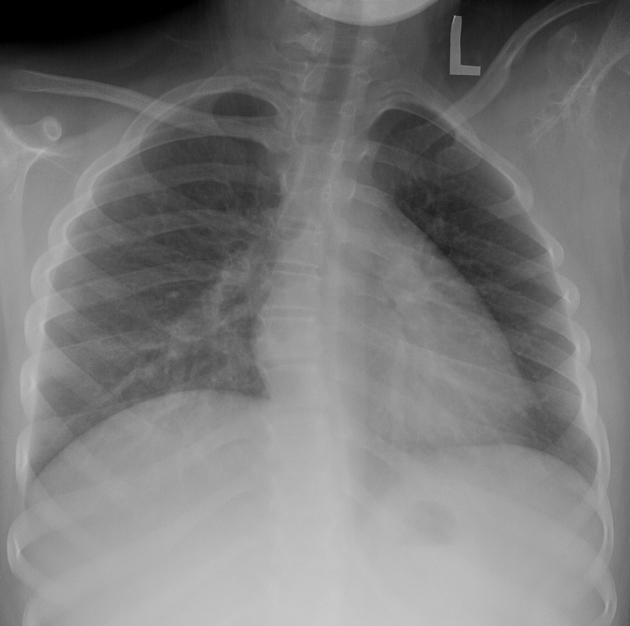
Chest, left shoulder and left humerus x-ray.
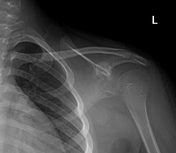
Asymmetric shoulders. Left shoulder is elevated and the scapula is displaced from the chest wall, especially on the chest x-ray. Associated soft-tissue mass extends into the axilla.
![](https://prod-images-static.radiopaedia.org/images/382573/downloaded_image20211020-1624-wg60dx_thumb.jpeg)
![This study is a stack](/packs/stack-YQKLCKBI.gif)
![](https://prod-images-static.radiopaedia.org/images/382604/downloaded_image20211020-1624-13tvuo6_thumb.jpeg)
![This study is a stack](/packs/stack-YQKLCKBI.gif)
![](https://prod-images-static.radiopaedia.org/images/382633/downloaded_image20211020-1627-lnl6nf_thumb.jpeg)
![This study is a stack](/packs/stack-YQKLCKBI.gif)
![](https://prod-images-static.radiopaedia.org/images/382663/downloaded_image20211020-1654-uqwusg_thumb.jpeg)
![This study is a stack](/packs/stack-YQKLCKBI.gif)
![](https://prod-images-static.radiopaedia.org/images/382573/downloaded_image20211020-1624-wg60dx_big_gallery.jpeg)
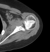
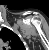
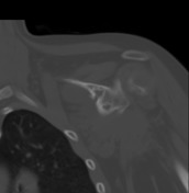
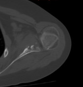
The abnormality of the left shoulder is centered on the subscapularis muscle. There is muscular expansion and adjacent bony destruction. In this age group, a primary muscle sarcoma is considered the most likely differential.
![](https://prod-images-static.radiopaedia.org/images/382700/a6371339ced3c1eb5878f7199d7f57_thumb.jpeg)
![This study is a stack](/packs/stack-YQKLCKBI.gif)
![](https://prod-images-static.radiopaedia.org/images/382717/10b8e417c41620834e45e35686f0bc_thumb.jpeg)
![This study is a stack](/packs/stack-YQKLCKBI.gif)
![](https://prod-images-static.radiopaedia.org/images/382740/a6b77130a77261e20437f2fbf34b51_thumb.jpeg)
![This study is a stack](/packs/stack-YQKLCKBI.gif)
![](https://prod-images-static.radiopaedia.org/images/382757/0300d6df594ad6258f89d136e32ec9_thumb.jpeg)
![This study is a stack](/packs/stack-YQKLCKBI.gif)
![](https://prod-images-static.radiopaedia.org/images/382772/b4e805a74e9344300ee2769678f2b7_thumb.jpeg)
![This study is a stack](/packs/stack-YQKLCKBI.gif)
![](https://prod-images-static.radiopaedia.org/images/382790/1b04dc2fd16af23ff431b2e52a8962_thumb.jpeg)
![This study is a stack](/packs/stack-YQKLCKBI.gif)
![](https://prod-images-static.radiopaedia.org/images/382700/a6371339ced3c1eb5878f7199d7f57_big_gallery.jpeg)
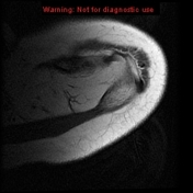
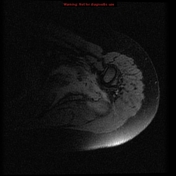
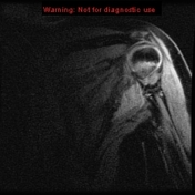
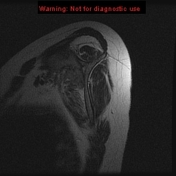
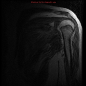
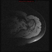
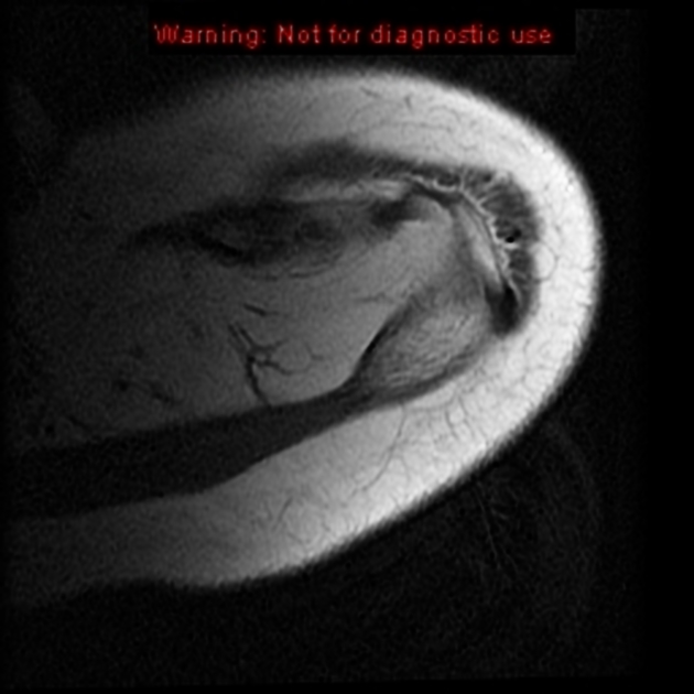
MRI confirms an irregular muscle-based tumor with bony destruction arising from subscapularis and extending into the axilla.
Case Discussion
The differential for this muscle-based tumor has sarcoma at the top (rhabdomyosarcoma is probably the most likely in this age group) with fibroblastic or myofibroblastic tumors in the differential. Biopsy was performed and this was pathologically proven fibromatosis.