
Presentation
Fever, chest pain. History of diabetes.
Patient Data
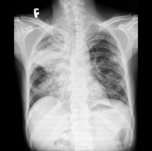
Right pulmonary consolidation with cavitation and a gas-fluid level in the upper lobe.
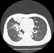
Multiple small nodules in both lungs with cavitation of some nodules
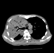

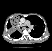
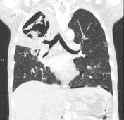
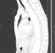
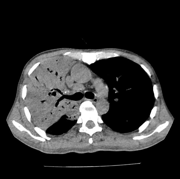
The consolidation is almost replacing the entire right upper lobe with air-bronchograms and cavitation. The consolidation also is seen in the right middle lobe and lower lobe.
There are multiple small solid nodules, ground-glass nodules, and tree-in-bud in both lobes with cavitation in some of the nodules.
Mediastinal lymph node enlargement.
Blood test: WBC 28G/l. Glucose 23mmol/l.
Blood culture: Burkholderia pseudomallei
Case Discussion
Melioidosis is an infectious disease caused by the bacterium Burkholderia pseudomallei. The lung is the most commonly affected organ.
This case demonstrates the right upper lobe pneumonia with cavity formation and multiple small pulmonary nodules consistent with the acute stage.


 Unable to process the form. Check for errors and try again.