
Presentation
Hematuria
Patient Data

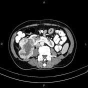
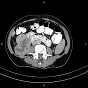
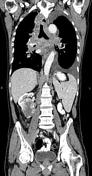

Left kidney is ectopic, located at midline with horizontal orientation and fused to the lower pole of right kidney. 95 x 65 mm heterogeneously enhancing lower pole right renal mass with central foci of necrosis. Multiple enlarged retroperitoneal nodes, e.g. paraaortic and paracaval regions, with short axis diameters less than 20 mm.
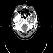
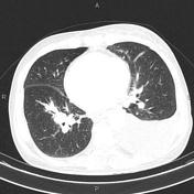
In addition, multilevel cervical nodal enlargement, left>right. The largest one measuring 37 × 23 mm. Multiple mediastinal and both hilar nodal enlargements. The largest one measuring 40 x 32 mm. There are also several small nodes of left axillary and retropectoral regions.
Left thyroid lobe 25 mm hypoattenuating nodule.
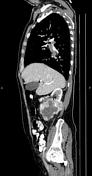
Moderate left-sided pleural effusion. Several atelectatic bands scattered bilaterally.
Prostate gland is enlarged.
Case Discussion
Right renal mass; pathology proven renal cell carcinoma (clear cell subtype) with retroperitoneal, mediastinal, hilar, axillary and cervical lymphadenopathies.


 Unable to process the form. Check for errors and try again.