
Presentation
Right cervical and facial palpable masses on physical exam.
Patient Data
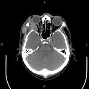


Enhancing tumoral lesion is present at the superolateral aspect of right orbit measuring 45×20×40 mm, causing lateral orbital wall bony destruction. A 38×17×30 mm extra orbital component is also evident. Apparent globe involvement is not seen.
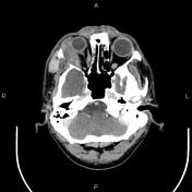
The lytic expansile lesion is noted at the posterolateral wall of the right maxillary sinus. Mucosal thickening is present at both maxillary sinuses, frontal sinus, and some ethmoidal air cells.
The right parotid gland is enlarged and shows inhomogeneous enhancement.
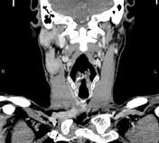
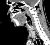
Several enlarged lymph nodes are seen in upper cervical regions with a maximum SAD of 28 mm.
Case Discussion
Pathology proved non-Hodgkin lymphoma with infiltration of the orbit, maxillary sinus walls, parotid gland, and cervical lymph nodes on the right side.


 Unable to process the form. Check for errors and try again.