
Presentation
Left flank pain and hematuria.
Patient Data
Age: 30 years
Gender: Female
From the case:
Renal cell carcinoma

Download
Info
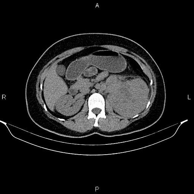
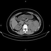
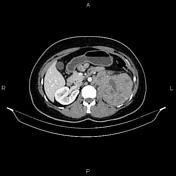
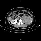
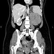
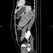
A 180 × 75 × 95 mm heterogeneously enhancing mass is noted at the left kidney. The left urinary collecting system and proximal portion of the left renal vein are obliterated by the mass. The fat plane between the mass and adjacent iliopsoas muscle is obliterated, and local invasion is suspected.
Several enlarged lymph nodes in a paraaortic location with a short-axis diameter less than 18 mm.
An IUD is present within the uterine cavity in the appropriate station.
Case Discussion
Huge left renal mass, pathology proved renal cell carcinoma (clear cell subtype) with local invasion, vascular extension, and regional lymphadenopathy.


 Unable to process the form. Check for errors and try again.