
Presentation
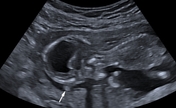
G2P1 at 16 weeks gestation. Routine ultrasound.
Patient Data
Age: 30 years
Gender: Female
From the case:
Alobar holoprosencephaly - antenatal


Download
Info
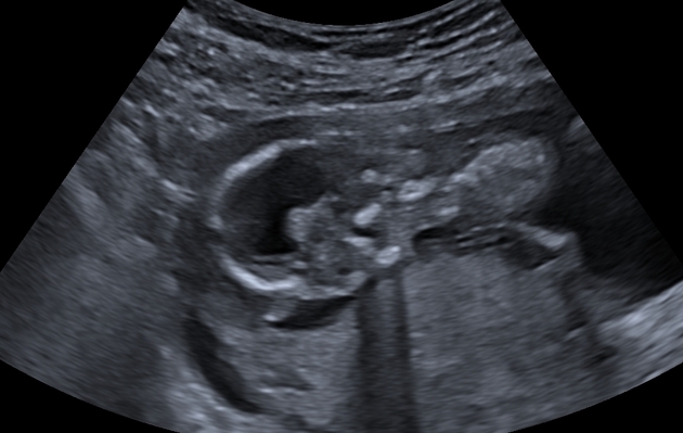
The ultrasound images demonstrate:
- the supratentorial cerebral parenchyma is replaced by a large central monoventricle with a thin rim of peripheral parenchyma
- fused thalami
- absent midline structures (septum pellucidum, corpus callosum, and falx cerebri)
- hypotelorism
- small midline structure of the fetal forehead suggestive of a proboscis
From the case:
Alobar holoprosencephaly - antenatal

Download
Info

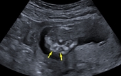
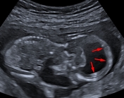
Annotated images:
- white arrow: small midline structure of the fetal forehead suggestive of a proboscis
- yellow arrow: hypotelorism
- red arrows: rim of cerebral cortex
- blue arrows: fused thalami
Case Discussion
Ultrasound findings most consistent with alobar holoprosencephaly
The main differential diagnosis includes:
-
hydranencephaly
- thalami usually visible and not fused
- falx cerebri usually present
- no associated midline facial abnormalities
- no cortex present, or sometimes small islands of tissue
- severe hydrocephalus
- thalami not fused
- no associated midline facial abnormalities
- falx cerebri usually present
- bilateral choroid plexus


 Unable to process the form. Check for errors and try again.