
Presentation
Work up for hematuria.
Patient Data
Age: 45 years
Gender: Female
From the case:
Renal cell carcinoma


Download
Info
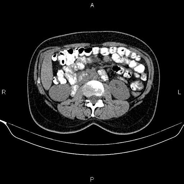
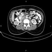
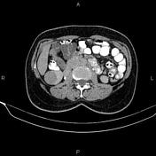
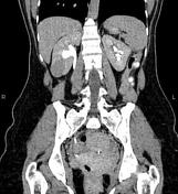
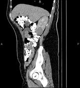
A 35 mm mass is seen in the lower pole of right kidney. The neoplasm has a similar density to normal renal parenchyma on non enhanced CT scan. After IV contrast media injection, the attenuation value increased from 31 HU to 102 HU. Nephrographic phase CT scan shows that the mass enhances less than normal renal parenchyma.
An 8 mm cyst is present at segment VIII of the liver.
An IUD is present within the uterine cavity in appropriate station.
Case Discussion
The patient underwent right partial nephrectomy and histopathologic evaluation confirms renal cell carcinoma (clear cell subtype).


 Unable to process the form. Check for errors and try again.