
Presentation
Large neck swelling since birth.
Patient Data


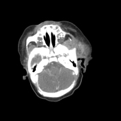

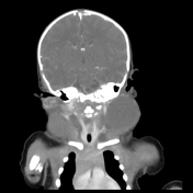
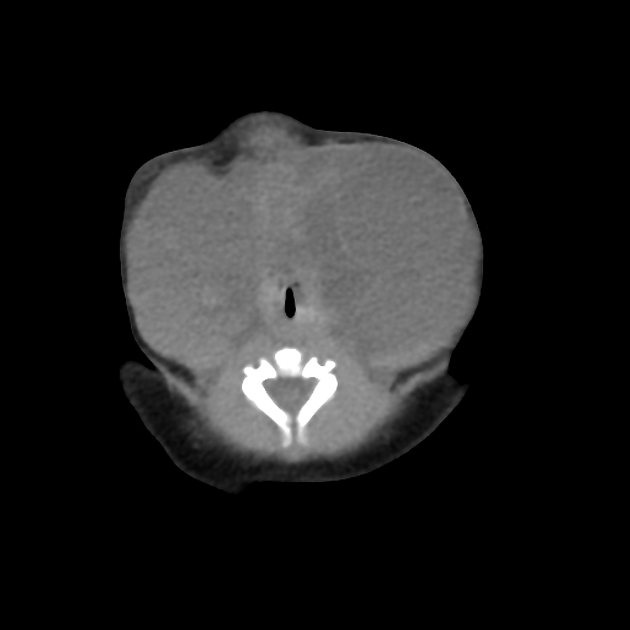
Bilateral cervicofacial sizable multilocular cystic lesions measure 6x5x6 cm and 7.5x5x7.5 cm at the right and left sides respectively. They extend caudally about the level of sternoclavicular joints with no mediastinal extension. They exert a bilateral symmetrical mass effect upon the airways as well as displacing and surrounding the neck vascular structures. They elicit fluid density with internal minimally enhanced septations.

Case Discussion
Here is a case of congenital cervicofacial swelling with CT features that are highly suggestive of cystic hygroma. They occur most commonly in the neck, which is then also termed nuchal cystic hygroma (occurs in ~80% of cases) and axilla, with only 10% of cases extending to the mediastinum and only 1% confined to the chest. This case shows no mediastinal extension.
The patient was prepared for lymphatic sclerotherapy.


 Unable to process the form. Check for errors and try again.