
Presentation
Work up for abdominal pain.
Patient Data
Age: 40 years
Gender: Male
From the case:
Chronic pancreatitis


Download
Info

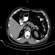
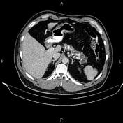
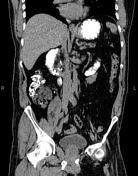
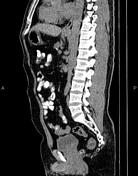
Coarse pancreatic calcifications are seen accompanied by duct dilatation, inferring chronic pancreatitis.
The right kidney is relatively smaller than the left kidney and shows parenchymal atrophic changes. A 38 mm stone is noted at right renal pelvis causing moderate hydronephrosis. A 30 mm stone is observed at left renal pelvis accompanied by mild hydronephrosis. Several small stones are also observed at lower calyces of the both kidneys.
The prostate gland is enlarged.
Case Discussion
CT features of chronic pancreatitis include dilatation of the main pancreatic duct, pancreatic calcification, changes in pancreatic size (i.e. atrophy), shape, and contour and pancreatic pseudocysts.


 Unable to process the form. Check for errors and try again.