
Presentation
Abnormal uterine bleeding.
Patient Data


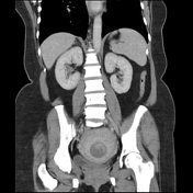
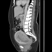

Large mass of the myometrium and endometrium involving >50% depth but no serosal breach or localized metastatic disease. No distal metastasis or pathological lymph node. Distended gallbladder with gallstones.

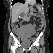
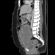
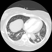
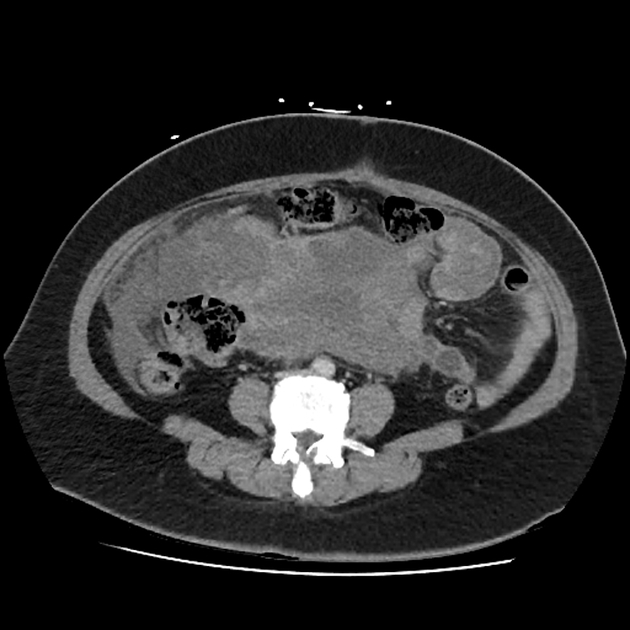
Impressive disease progression with diffuse pulmonary metastatic disease and diffuse abdominal involvement.
Case Discussion
This is a case of a 50-year old lady that presented with abnormal uterine bleeding to the gynecology outpatients department. Unfortunately, there was a delay to theater given patient non-compliance. A repeat CT was performed 4 months after the first CT and showed impressive progressive disease.
The patient eventually underwent a total abdominal hysterectomy and bilateral salpingo-oophorectomy with omental biopsies. Intra-op findings:
- disease eroding through serosa with disease on the omentum
- normal ovaries and peritoneal surfaces
Histopathology results: Uterine leiomyosarcoma, high grade, no lymphovascular invasion, metastatic disease to omentum, surgical margins adequate.


 Unable to process the form. Check for errors and try again.