
Presentation
Work up for abdominal pain and progressive distention.
Patient Data


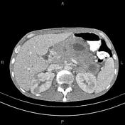
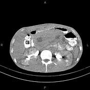
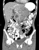
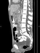
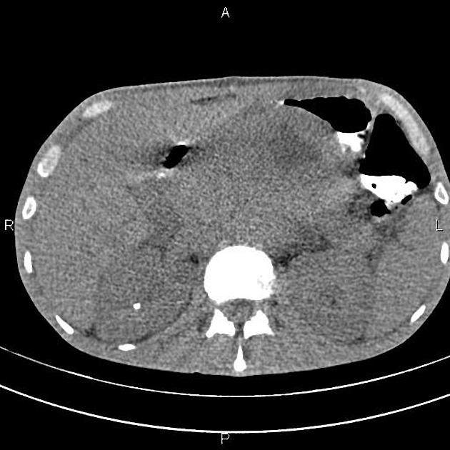
A 135×70 mm mass with internal necrosis is present at the anatomical location of the pancreas, which encases adjacent vascular structures.
Multiple small ill defined masses are seen at the liver less than 22 mm.
Both adrenal glands are infiltrated and enlarged.
There are also multiple well defined low enhancing masses in both kidneys.
Some abdominopelvic free fluid is evident.
A 4 mm stone is noted at the mid portion of the right kidney.
In imaged portions of the lower thorax, a pleural effusion is present bilaterally right more than left. A few nodules are seen at both lung fields less than 10 mm. Mild to moderate pericardial effusion is also observed.
Case Discussion
Pathology proved case of muli organ extranodal non-Hodgkin lymphoma.


 Unable to process the form. Check for errors and try again.