
Presentation
Epilepsy.
Patient Data
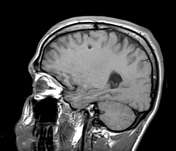

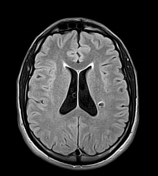
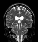

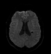
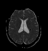

Numerous small well-defined intra-axial lesions in both cerebral hemispheres, the largest lesion (diametre = 10 mm) is located in the left paraventricular region. They elicit a low signal on T1 and T2 with prominent blooming on the GE sequence. The largest lesion shows surrounding edema of high signal on T2 and FLAIR indicating most likely a recent bleed. No enhancement on the post-contrast sequence.
The right ocular globe is small in size with a vitreous hemorrhage (known).
Case Discussion
MRI features of multiple small cerebral cavernous malformations (or cavernomas).
Cerebral cavernous venous malformations or cavernomas are the third most common cerebral vascular malformation after developmental venous anomaly and capillary telangiectasia.
Most patients have a single lesion. Multiple lesions may be familial and screening of family members may be indicated.


 Unable to process the form. Check for errors and try again.