
Presentation
Abdominal pain.
Patient Data


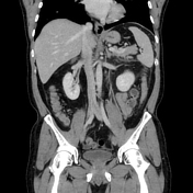

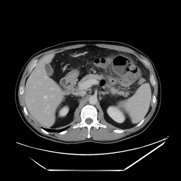
Focal walled-off outpouching of the proximal jejunum with a clear communicating thin channel with the lumen. Surrounding inflammation and localized peritonitis. Reactive inflammation of the distal transverse colon as well. Few other small, noninflamed jejunal diverticula (review coronal images).
Case Discussion
Typical findings of small bowel diverticulitis, which often looks like an abscess at first glance. Certainly in this case there is a lot of surrounding inflammation resulting in peritonitis and reactive thickening of the adjacent transverse colon and jejunal loops. However, you can be confident about this diagnosis by looking for a channel of communication with the small bowel (clearly seen on axial images in this case), and also other small bowel diverticula, which are often best seen on coronal images. It is also helpful to keep in mind that small bowel diverticula get quite enlarged when inflamed, compared to the relatively small colonic diverticula we become accustomed to.


 Unable to process the form. Check for errors and try again.