
Presentation
Cough and febrile symptoms. No improvement despite antibiotics.
Patient Data
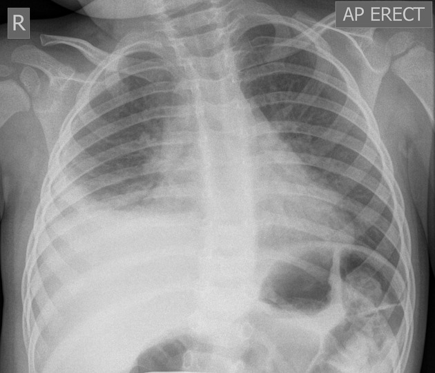
Dense consolidation in the right lower zone with an adjacent basal pleural effusion. Background moderate perihilar, peribronchial thickening is consistent with airway inflammation. The left lung is clear of consolidation.
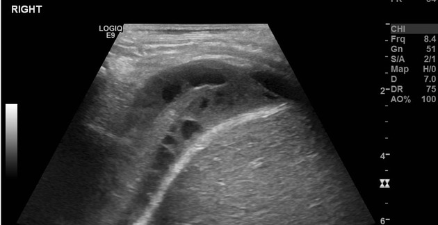
Targeted ultrasound examination of the right hemithorax demonstrates a complex collection in the pleural space. The collection has heterogenous internal echoes and septations, without vascularity, consistent with empyema.
The adjacent right basal lung is consolidated/collapsed.
Case Discussion
Pleural empyemas usually occur secondary to underlying pneumonia, with pneumococcus the most common cause in pediatric patients. Empyema may be suspected if there is the failure of resolution of infectious/febrile symptoms in a child with pneumonia and a persistent pleural effusion on a chest x-ray.
If available, ultrasound evaluation of pleural fluid collections is usually preferred over CT as a first-line modality in pediatric patients.
While this case demonstrates typical features for pleural empyema. A pleural fluid infection cannot be excluded on imaging alone even if the effusion appears simple on ultrasound.


 Unable to process the form. Check for errors and try again.