
Metastatic renal cell carcinoma with synchronous urinary bladder transitional cell carcinoma
Presentation
Work up for gross hematuria.
Patient Data

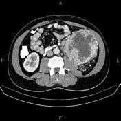
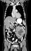
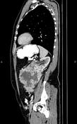

A 165×120×125 mm exophytic hetero enhancing mass with areas of internal necrosis is present at the lower pole of the left kidney without signs of vascular extension.
Several para-aortic lymphadenopathies are noted with SAD less than 23 mm.
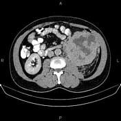
Increased wall thickness up to 12 mm is present at the dome and left lateral aspect of the urinary bladder, which is suggestive of tumor infiltration.
Several hypervascular masses are seen at liver less than 20 mm that become isodense with surrounding parenchyma on delayed images.
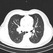
Several nodules are seen at both lungs less than 10 mm.
A few enlarged lymph nodes are seen at hilar regions bilaterally.
The prostate gland is enlarged.
Case Discussion
The patient underwent a left nephrectomy, and histopathology evaluation confirms renal cell carcinoma (clear cell type).
In addition, cystoscopy was performed for the patient, and the biopsy showed urothelial cell carcinoma.


 Unable to process the form. Check for errors and try again.