
Presentation
Left clavicular region nodule for the last 18 months, gradually increasing in size, eliciting mild local pain.
Patient Data
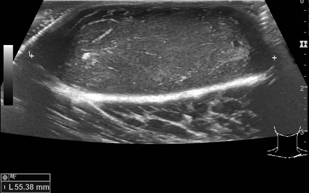
There is a well-defined subcutaneous lesion in the left clavicular region measuring about 55 x 44 x 20 mm. It exhibits linear and amorphous echogenic areas without any calcification or cystic changes. The lesion shows acoustic enhancement. Few internal flow signals are present. There is no extension to the underlying muscle and the adjacent clavicular cortex does not show erosion.
Case Discussion
A young male presented with a gradually increasing anterior chest wall lesion along with mild local pain. The ultrasound showed a solid subcutaneous lesion. Surgical excision was performed. The histopathology diagnosis was of a dermatofibrosarcoma protuberans.


 Unable to process the form. Check for errors and try again.