
Presentation
Shortness of breath, cough for 4 months but no constitutional symptoms
Patient Data

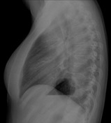
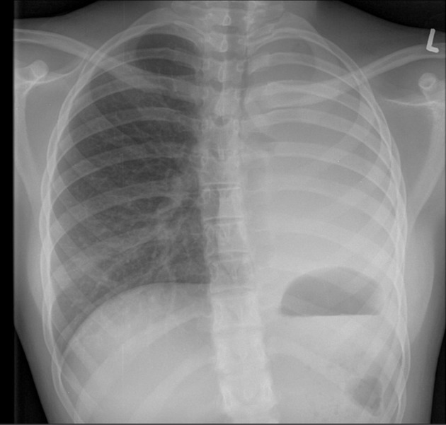
Left hemithorax volume loss with a tracheal shift to the left, elevated gastric bubble and rib crowding. Non-visualization of left hemidiaphragm outline with complete white out of the left lung and abrupt cut-off of the left mainstem bronchus. No calcification or rib destruction was noted.


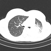
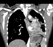
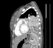

Large avidly enhancing endobronchial mass in the proximal left mainstem bronchus with distal atelectasis and hypodense bronchoceles distally. The centrally located mass has no cavitation or foci of calcification. Associated left hemithorax volume loss as evidenced by mediastinal shift, the elevation of hemidiaphragm and rib crowding. Small left pleural effusion. No hepatic or adrenal lesion.


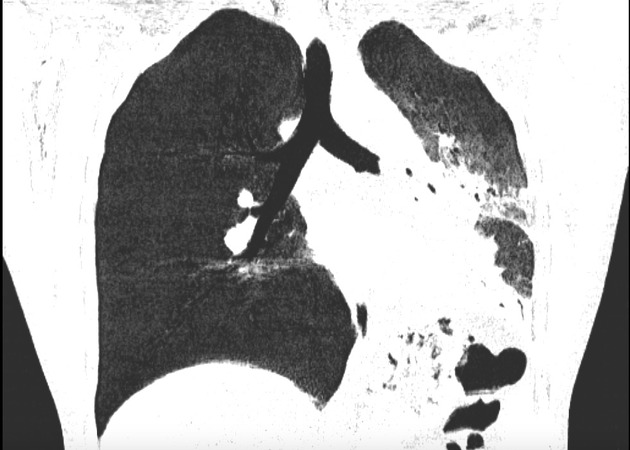
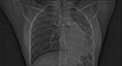
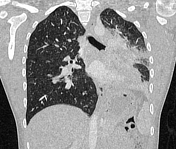
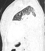
Minimum intensity projection (MinIP) axial and coronal view images showing the endobronchial lesion with an abrupt truncation of the left mainstem bronchus, distal atelectasis and mediastinal shift to the left.
Case Discussion
The patient had a surgical lobectomy and histopathological confirmation of a typical carcinoid tumor. Differential diagnoses would include hypervascular endobronchial metastasis which is rare.


 Unable to process the form. Check for errors and try again.