
Presentation
Low back pain radiating down to the legs (more on the left side), associated with numbness in the lower limbs for one year. No weakness, paralysis or sphincter disturbance.
Patient Data


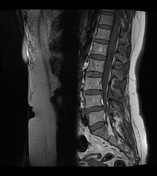
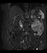
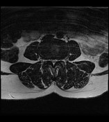
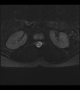
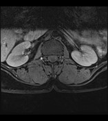
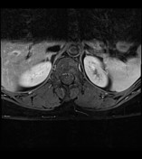
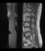
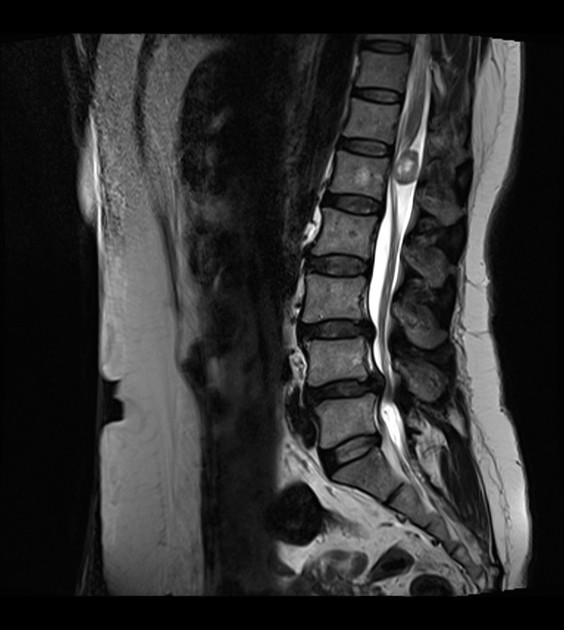
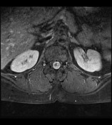
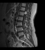
Well-defined oval shaped intradural extramedullary nodule measuring 14 x 13 mm at the level of conus medullaris. It is isointense on T1, mixed signal on T2-weighted images and shows moderate enhancement on the post-contrast study. A few small non-enhancing cystic areas are seen within it.
Small osseous hemangioma in the 1st lumbar vertebral body. Disc protrusions at L3-L4 and L4-L5 levels causing indentation on thecal sac and cauda equina. A few small focal hyperintense lesions in the liver which are hepatic cysts and a few small hypointense lesions in the uterus which are likely fibroids.

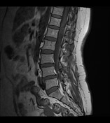
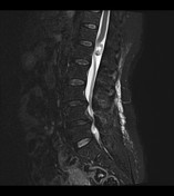

Stable intradural extramedullary lesion at the thoracolumbar junction just below the conus medullaris.
Small well-defined moderately enhancing intradural extramedullary lesion, which is likely a neurofibroma or schwannoma.
Case Discussion
The patient underwent laminectomy and excision of the lesion.
Histopathology: Schwannoma.


 Unable to process the form. Check for errors and try again.