
Presentation
Right upper quadrant pain and fullness on physical exam.
Patient Data
Age: 40 years
Gender: Male
From the case:
Thoracoabdominal hydatid cysts


Download
Info
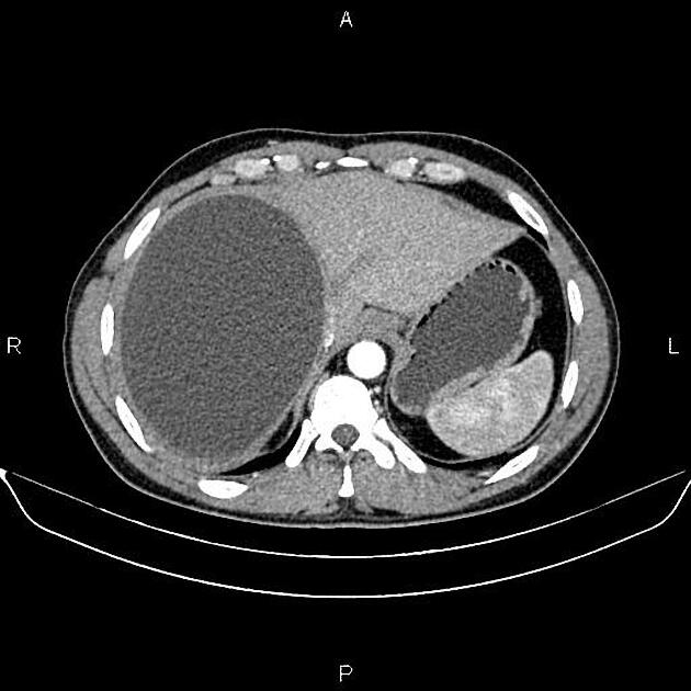
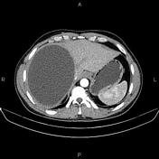
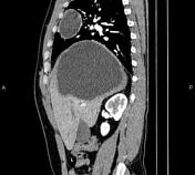
A 165×120×130 mm unilocular thick walled cystic lesion is noted at the right liver lobe, which causes right hemidiaphragm elevation. No calcification or enhancing solid components are pointed out within.
In imaged portions of the lower thorax, a 58×46 mm cystic lesion with a similar appearance is also noted at the anteromedial aspect of the right lung.
Case Discussion
Features on CT images are most consistent with thoracoabdominal hydatid cysts (serologic correlation).


 Unable to process the form. Check for errors and try again.