
Presentation
Central chest pain and shortness of breath for 3 months
Patient Data

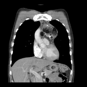
CT scout view shows a mediastinal mass projected over the left hilar region with the aortic contour preserved and the hilum overlay sign present, consistent with an anterior mediastinal mass.
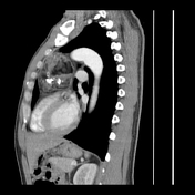

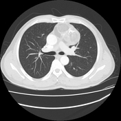

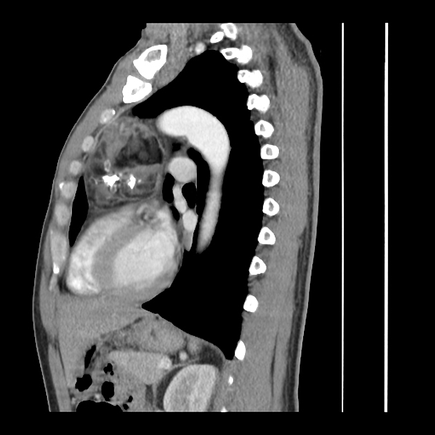
Large well-circumscribed anterior mediastinal mass measuring 85 mm TRV x 70 mm AP x 80 mm CC.
The mass is heterogeneous with macroscopic fat, soft tissue density and coarse calcifications.
Subcentimeter mediastinal lymph nodes. No hilar lymphadenopathy.
Case Discussion
Mediastinal teratomas are rare extragonadal germ cell tumors. The majority are asymptomatic, but in one series of 66 patients 1, 70% of patients presented with symptoms, predominantly chest pain, dyspnea and cough.
The case demonstrates a good example of the hilum overlay sign, described by Benjamin Felson, which states that a mediastinal mass anterior or posterior to the hilum will often extend laterally for a considerable distance and result in preserved visualization of the hilar vessels which can often be tracked medially to their confluence 2. Computed tomography (CT) with intravenous contrast is the most important radiologic tool for evaluation since it depicts different tissue attenuations 3. A teratoma will typically be cystic and contain varying amounts of fat, soft tissue and calcification as in this particular case.


 Unable to process the form. Check for errors and try again.