
Presentation
Abdominal pain.
Patient Data


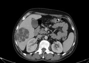
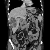
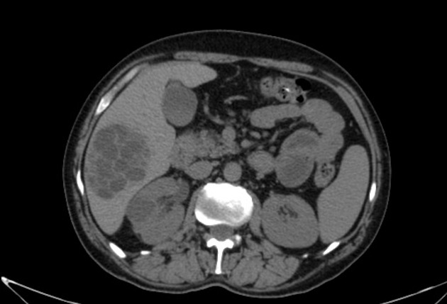
Right hepatic lobe well-defined lesion with fluid attenuation and multiple daughter cysts inside giving honeycomb/rosette pattern. There is no calcification or any enhancing components within.
Moderate splenomegaly and multiple enlarged reactive upper abdominal lymph nodes are also detected.
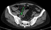
Minimal right-sided hydroureteronephrosis down to a minute hyperdense stone seen within the right pelvic ureter (green arrow in axial non-contrast image) and a few bilateral renal simple cortical cysts are also noted.
Case Discussion
This patient is a shepherd. CT findings along with the patient history are consistent with hepatic hydatid cyst. The liver is the most common site of hydatid infection.
Honeycomb / Rosette pattern is a very characteristic sign, which helps in diagnosing hydatid cysts. It represents multivesicular mother cyst with daughter cysts separated by radiating septae representing cyst walls and hydatid sand/matrix.
Right calcular obstructive uropathy was also there.


 Unable to process the form. Check for errors and try again.