
Presentation
History of resected choroid plexus papilloma
Patient Data
Age: Adult
Note: This case has been tagged as "legacy" as it no longer meets image preparation and/or other case publication guidelines.
From the case:
Spinal cord metastasis

Download
Info
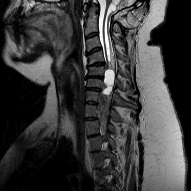
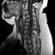
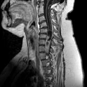
An intramedullary complex lesion is noted opposite the lower cervical and upper dorsal vertebrae. It shows isointense signal on T1, high T2 signal and homogeneous postcontrast enhancement. It is compressing central spinal canal with proximal syrinx. It shows cystic component with signal similar to CSF.
Case Discussion
Patient with a choroid plexus papilloma previously resected (note the posterior fossa craniectomy) subsequently developed a spinal cord metastasis.


 Unable to process the form. Check for errors and try again.