
Presentation
Work up for abdominal pain.
Patient Data
Age: 85 years
Gender: Female
From the case:
Hydatid cysts


Download
Info
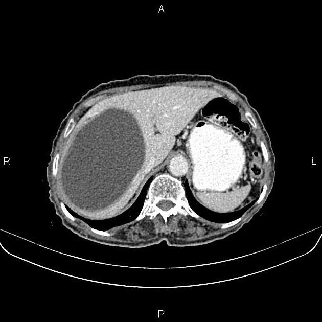
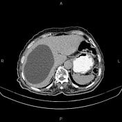
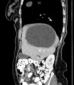
A 115×100×95 mm thick walled cystic lesion is noted at the right liver lobe without apparent enhancing solid component or calcification. Mild adjacent intrahepatic bile ducts dilatation is evident most likely due to the pressure effect.
In addition, several small cystic lesions are seen in the porta hepatis and peripancreatic regions less than 20 mm in diameter.
In imaged portions of the lower thorax; a 25×18 mm cystic lesion is noted at the right lung.
Case Discussion
Hepatic, peritoneal, and pulmonary cystic lesions; consistent with hydatid cysts, confirmed with serology.


 Unable to process the form. Check for errors and try again.