
Presentation
Pain and swelling at medial aspect of wrist
Patient Data


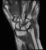
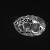
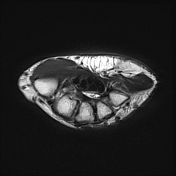
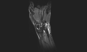
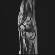
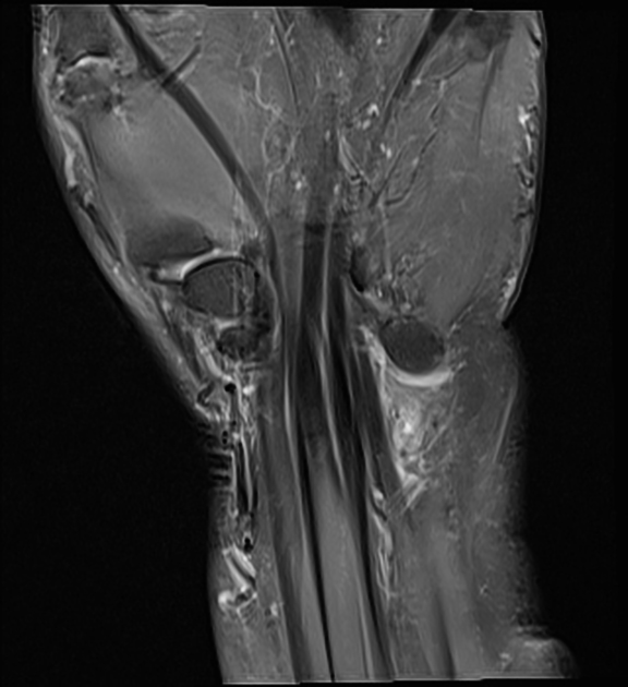
Significant fluid along extensor carpi ulnaris tendon with distension of its sheath and areas of synovial thickening, involving a length of approx. 55 mm starting from the distal forearm up to its insertion at the base of the fifth metacarpal.
Mild thickening and hyperintensity involving extensor carpi ulnar tendon is also seen with possible small partial interstitial tear involving approx. 5 mm long segment near its insertion.
Significant radiocarpal joint effusion is seen with areas of synovial thickening.
TFCC: Partial tears of foveal attachment of triangular fibrocartilage is seen with low to intermediate grade sprain of styloid attachment and associated small erosions, mild marrow edema in distal ulna. Sprain of dorsal and volar radioulnar ligaments is seen with possible partial tears of ulnar attachment. Partial tears of ulnomeniscal homologue are also seen.
Case Discussion
Findings reflecting extensor carpi ulnaris tenosynovitis with a possible small partial interstitial tear. These explain the patient's symptoms of swelling and pain.


 Unable to process the form. Check for errors and try again.