
Presentation
Diplopia.
Patient Data
Age: Young adult
Gender: Male
From the case:
Cavernous malformation: midbrain

Download
Info
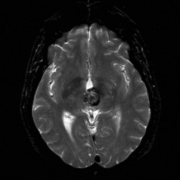
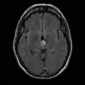
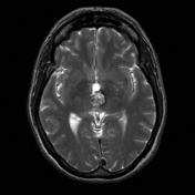
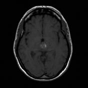
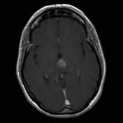
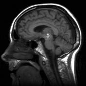
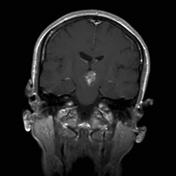
Centered in the superior midbrain is a rounded lesion with peripheral signal drop-out (best seen on gradient echo sequence - GRE) consistent with hemosiderin. Centrally the lesion is markedly heterogeneous in signal with regions of high signal on both T1 and T2 weighted images. There is no peripheral edema and no evidence of hydrocephalus.
Features are characteristic of a midbrain cavernous malformation.
Case Discussion
A cerebral cavernous hemangioma is a common type of cerebral vascular malformation. MRI is the modality of choice, demonstrating a characteristic “popcorn” or "berry" appearance with a rim of signal loss due to hemosiderin.


 Unable to process the form. Check for errors and try again.