
Abdominal aortic aneurysm with intramural hematoma and ulcer-like projection then rupture
Presentation
Abdominal pain. BP stable. Known AAA.
Patient Data
Initial CT


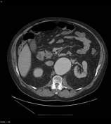

The non contrast phase demonstrates a bilobed suprarenal AAA extending to the bifurcation with some subtle peripheral hyperdensity in the lower lobe forming a crescent concerning for intramural hematoma. There is some mild left retroperitoneal stranding.
The CTA demonstrates eccentrically thickened aortic wall in the lower lobe of the aneurysm. No focal contrast blush is seen outside the aorta. The kidneys appear well perfused.
The patient was transferred to a tertiary center and started to become hypotensive but responsive to packed red cells and fluids. A repeat CTA was arranged upon arrival at the tertiary center.
11 hrs later - transfer to...
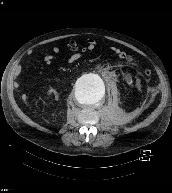
11 hrs later - transfer to tertiary center
There is now more left retroperitoneal stranding and fluid which is displacing the left kidney anteriorly suggesting ongoing slow rupture. In the left posterolateral wall of the lower lobe of the bilobed AAA, there is contrast now extending into the wall in keeping with progression of the acute intramural hematoma (development of an ulcer-like projection (ULP)). This is at the site of the wall thickening and crescent sign on the initial CT.
No active contrast blush is seen outside the aorta. Again, the kidneys appear well perfused.
Case Discussion
The patient proceeded to surgery urgently and an open AAA repair was performed. The surgical notes describe only a thin layer of adventitia lining the intramural hematoma, with frank rupture only 'seconds away'.


 Unable to process the form. Check for errors and try again.