
Presentation
Epigastric pain.
Patient Data
Age: 70 years
Gender: Female
From the case:
Abdominal foreign body - fish bone


Download
Info
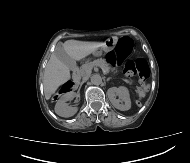
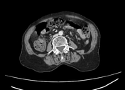
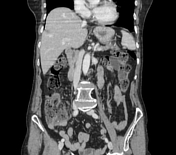
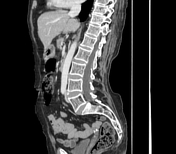
The CT images demonstrate a linear hyperdense structure in the posterior wall of the antral region, extending to the retrogastric space surrounded by an enhancing inflammatory tissue with fat stranding.
Case Discussion
The clinical history is suggestive of an ingested fish bone causing transmural perforation of the posterior antral wall with surrounding inflammatory granuloma. The upper GI endoscopy confirms the gastric perforation.


 Unable to process the form. Check for errors and try again.