
Presentation
Sudden sharp pain with audible snap in the back of the right ankle and inability to walk correctly while football.
Patient Data


-
Achilles tendon show;
- Is thicker than normal (swelling to >12 mm AP) and has a convex anterior surface, with discontinuity of most of its fibers, but still minor intact fibers.
- The torn area is 5 cm above its insertion in the calcaneus with a gap of about 1.3 mm.
- Organized hematoma in place of the normal tendon.
- The tear has high signal intensity on fluid-sensitive sequences.
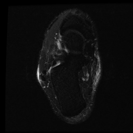
- Note the intact plantaris tendon inserted into the medial tuber calcanei.
- Subcutaneous edema at the posterolateral aspect of the ankle joint.
- Mild ankle joint effusion.
OPINION:
The MRI findings suggest a complete Achilles tendon tear (Type II) according to Kuwada classification of Achilles tendon tear with an overlap of its edges, hematoma, fluid and tissues filling the gap in between.


Sagittal PD fat-suppressed and sagittal T1 weighted images demonstrate a proximal Achilles tendon tear extending to the anterior surface of the Achilles tendon with thickening and heterogeneity of the Achilles tendon bordering the tear (red arrow) at the myotendinous junction with a small gap (green arrow ).
A sagittal PD and axial T1 fat-suppressed images demonstrate an intact Plantaris tendon (blue arrows) inserted into the medial tuber calcanei.
Case Discussion
A 25-year-old male with a typical presentation of a sudden onset of pain at the back of the heel. (typical tear of the Achilles tendon while playing sports). It is typically located at the critical zone, usually approximately 5 cm above its insertion. MRI demonstrate a proximal Achilles tendon tear at the myotendinous junction with a small gap. The patient underwent surgical repair and confirmed the diagnosis of a complete torn Achilles tendon.
The diagnosis of Achilles tear is largely clinical but MRI is valuable and confirming for evaluating patients requiring surgical intervention or who do need conservative measures.


 Unable to process the form. Check for errors and try again.