
Presentation
Right iliac fossa abdominal pain, mild fever, and blood tests show elevated white blood cell count and increased percentage of neutrophils.
Patient Data
Age: 25 years
Gender: Male
From the case:
Acute appendicitis


Show annotations
Download
Info

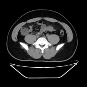
On CT, the distal appendix is noted to be enlarged, measuring approximately 10mm in diameter, with thickened walls enhancing after contrast administration, and a appendicolith present in the lumen. Surrounding fat stranding and thickening of the adjacent peritoneum are observed. Diffuse hepatic steatosis is also noted.
Case Discussion
The abdominal CT findings are consistent with acute appendicitis in the right iliac fossa. The patient subsequently underwent laparoscopic surgery, which confirmed the diagnosis, and the appendix was removed.


 Unable to process the form. Check for errors and try again.