
Presentation
Fever and right iliac fossa pain
Patient Data


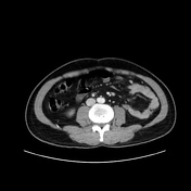
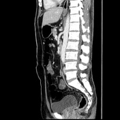
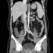

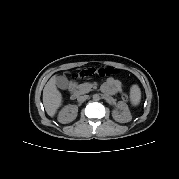
The appendix is enlarged measuring about 12 mm with thickened wall and increased post-contrast enhancement associated with mild stranding of periappendiceal fat.
Incidentally noted circumaortic left renal vein.
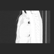
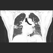
Bilateral multifocal peripheral ill-defined ground-glass opacities with basal and posterior predominance, associated with few subpleural atelectatic bands.
Case Discussion
The patient presented with right iliac fossa pain and fever, no respiratory manifestations, so the clinical diagnosis was acute appendicitis. Routine chest X-ray was suspicious of COVID 19 pneumonia. Therefore, CT chest, abdomen and pelvis was done and revealed acute appendicitis and typical CT features of COVID 19 pneumonia. The first nasopharyngeal swab came negative; however, the second one was positive.


 Unable to process the form. Check for errors and try again.