
Presentation
Vague abdominal pain for 1 week. Increased pain in upper abdomen for 1 day. Suspected pancreatitis.
Patient Data

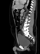
An inflamed diverticulum (coronal, axial) is seen in the sigmoid colon with surrounding fat streakiness.
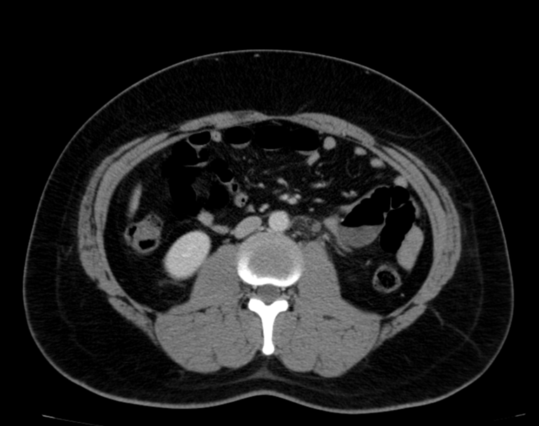
A few diverticula are also noted at the anterior aspect of the sigmoid colon.
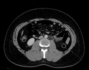
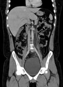
The inferior mesenteric vein (IMV) shows a lack of contrast enhancement within its lumen, associated with edema of the adjacent fat planes along its entire course, suggesting IMV thrombosis (axial, coronal).
The liver demonstrates apparent filling defects in the distal right portal vein branches, periportal edema, and heterogeneous contrast enhancement in the porto-venous phase.
Wall thickening of the gallbladder is noted.
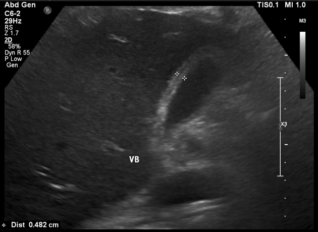
Ultrasound performed four hours after CT showed thickening of the gallbladder wall without intraluminal stones. No remarkable findings in the liver.
Case Discussion
Findings are consistent with acute diverticulitis (Hinchey stage Ia). Additionally, there is evidence of septic thrombophlebitis of the inferior mesenteric vein. The liver demonstrates a geographic pattern with apparent filling defects in the distal portal vein branches, periportal edema, and thickening of the gallbladder wall. Hematogenous dissemination from the pelvic infectious focus and associated pylephlebitis are suspected.


 Unable to process the form. Check for errors and try again.